Home > Popular Themes > Human Body
HeLa cells, light micrograph C017 / 8298

![]()
Wall Art and Photo Gifts from Science Photo Library
HeLa cells, light micrograph C017 / 8298
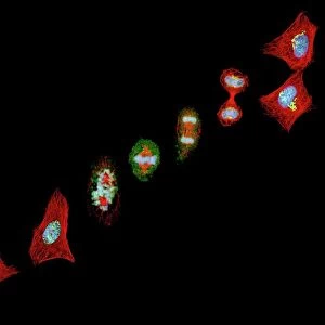
HeLa cells, multiphoton fluorescence micrograph (MFM). The cell nuclei, which contain the cells genetic information, are blue. Golgi bodies, which modify and package proteins, are orange. Microtubules, protein filaments that make up part of the cytoskeleton, are green. The cytoskeleton maintains the cells shape, allows some cellular mobility and is involved in intracellular transport. HeLa cells are a continuously cultured cell line of human cancer cells. They are immortal and so thrive in the laboratory. HeLa cells are widely used in biological and medical research
Science Photo Library features Science and Medical images including photos and illustrations
Media ID 9211211
© NATIONAL INSTITUTES OF HEALTH/SCIENCE PHOTO LIBRARY
Cancer Cell Cervical Cervix Culture Cultured Cytological Cytology Cytoskeletal Cytoskeleton Filament Filaments Fluorescence Fluorescent Golgi Apparatus Golgi Body Hela Cell Immortal Immunofluorescent Malignant Micro Organism Microbe Microbiology Microorganism Microtubule Microtubules Nuclei Nucleus Oncology Organelle Abnormal Cells Light Micrograph Light Microscope Protein
EDITORS COMMENTS
This print showcases HeLa cells, a remarkable and extensively studied cell line derived from human cancer cells. The image, captured using a light microscope, reveals the intricate details of these immortal cells that thrive in laboratory settings. The blue hues highlight the cell nuclei, which house the genetic information crucial for cellular functions. In contrast, the vibrant orange color represents Golgi bodies – organelles responsible for modifying and packaging proteins within the cell. These proteins play vital roles in various biological processes. Green microtubules are another prominent feature depicted in this photograph. Composed of protein filaments, they form part of the cytoskeleton – a network that maintains cell shape and enables mobility while facilitating intracellular transport. HeLa cells have revolutionized both biological and medical research due to their unique properties and ability to continuously divide outside their natural environment. Their malignant nature makes them particularly valuable for studying cancer-related phenomena. This visually striking image not only offers insights into cytology but also serves as a reminder of the immense impact HeLa cells have had on fields such as oncology, immunofluorescent studies, and microbiology. It captures an extraordinary moment frozen in time – a testament to humanity's relentless pursuit of understanding life at its most fundamental level. Photo credit: NATIONAL INSTITUTES OF HEALTH/SCIENCE PHOTO LIBRARY
MADE IN THE USA
Safe Shipping with 30 Day Money Back Guarantee
FREE PERSONALISATION*
We are proud to offer a range of customisation features including Personalised Captions, Color Filters and Picture Zoom Tools
SECURE PAYMENTS
We happily accept a wide range of payment options so you can pay for the things you need in the way that is most convenient for you
* Options may vary by product and licensing agreement. Zoomed Pictures can be adjusted in the Cart.