Human anatomy, 1823 C017 / 8057

![]()
Wall Art and Photo Gifts from Science Photo Library
Human anatomy, 1823 C017 / 8057
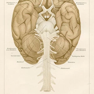
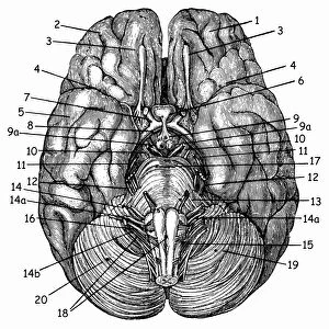
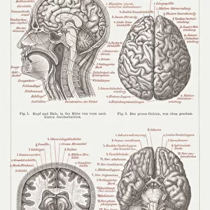
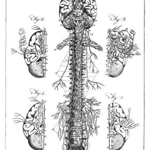
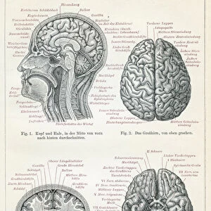
Human anatomy. 19th-century illustrations showing the anatomy of a human skull (upper left), brain and spinal column (upper centre), chest and abdominal organs (lungs, heart, liver, stomach, intestines; upper right), the heart (centre left), the pancreas (centre right), the urinary system (kidneys and bladder; lower left), lung airways (lower centre), and the stomach and small and large intestines (lower right). Details include spinal nerves and vertebrae, lung blood vessels either side of the heart, the pancreatic duct, and blood vessels adjacent to the kidneys. This page is from Universal Technological Dictionary (1823) by British author George Crabb (1778-1851)
Science Photo Library features Science and Medical images including photos and illustrations
Media ID 9210537
© MIDDLE TEMPLE LIBRARY/SCIENCE PHOTO LIBRARY
1823 Abdomen Abdominal Array Artworks Bladder Blood Vessels Bones Book Cardiac Chest Cranium Diagram Diagrams Digestive George Crabb Illustrations Intestines Kidneys Large Intestines Liver Lungs Organs Page Pancreas Pancreatic Duct Physiological Physiology Publication Respiratory Selection Small Intestines Spinal Column Spinal Nerves Stomach System Systems Thoracic Universal Technological Dictionary Urinary System Vascular Vertebral Column Brain Vertebrae
EDITORS COMMENTS
This 19th-century illustration, titled "Human Anatomy, 1823" offers a fascinating glimpse into the intricate workings of the human body. The print showcases various anatomical structures with meticulous detail and precision. In the upper left corner, we see a depiction of the human skull, highlighting its complex composition. Moving to the upper center, our attention is drawn to the brain and spinal column, showcasing their vital role in our nervous system. The upper right portion of this artwork presents an array of chest and abdominal organs such as lungs, heart, liver, stomach, and intestines. These essential components work harmoniously to sustain life within us. As we shift our focus towards the center left section of this illustration, we encounter a detailed representation of the heart - an organ synonymous with vitality and emotion. Continuing our exploration through this remarkable piece of scientific artistry brings us to other crucial systems within our bodies. The lower left area reveals the urinary system consisting of kidneys and bladder while moving downwards exposes lung airways in all their intricacy. Finally reaching the lower right corner unveils another set of vital organs: stomach and small/large intestines responsible for digestion and nutrient absorption. Throughout this masterpiece by British author George Crabb from his publication Universal Technological Dictionary (1823), one cannot help but marvel at each physiological structure's complexity. This monochrome artwork serves as both an educational tool for understanding human anatomy during that era as well as a testament to Crabb's dedication in disseminating knowledge about biology in his time.
MADE IN THE USA
Safe Shipping with 30 Day Money Back Guarantee
FREE PERSONALISATION*
We are proud to offer a range of customisation features including Personalised Captions, Color Filters and Picture Zoom Tools
SECURE PAYMENTS
We happily accept a wide range of payment options so you can pay for the things you need in the way that is most convenient for you
* Options may vary by product and licensing agreement. Zoomed Pictures can be adjusted in the Cart.