Vaccinia virus infected cell

![]()
Wall Art and Photo Gifts from Science Photo Library
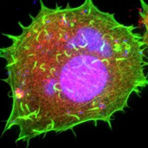
Vaccinia virus infected cell
Vaccinia virus infected cell. Immunofluorescence deconvolution micrograph of a cell infected with vaccinia virus particles. Viral DNA (deoxyribonucleic acid) is blue and its presence highlights areas of virus assembly within the cell. Actin protein filaments, which make up part of the cytoskeleton, are green. The cytoskeleton maintains the cells shape and is involved in intracellular transport. The virus uses the actin to propel the newly formed particles out of the cell, forming the protrusions seen here. The tyrosine kinase c-Abl, which is involved in catalysing actin motility, is red
Science Photo Library features Science and Medical images including photos and illustrations
Media ID 6308615
© DR DAN KALMAN/SCIENCE PHOTO LIBRARY
Actin Assembly Confocal Micrograph Cowpox Virus Cytoskeleton Deconvolution Filament Filaments Fluorescent Immunofluorescence Immunofluorescent Infected Infection Microtubules Nucleus Protrusion Protrusions Re Production Replicating Replication Reproducing Stain Stained Vaccinia Viral Virology Bio Chemistry Biochemical Deoxyribonucleic Acid Light Micrograph Light Microscope Micro Biology Microbiological Pathogen Protein Virus
MADE IN THE USA
Safe Shipping with 30 Day Money Back Guarantee
FREE PERSONALISATION*
We are proud to offer a range of customisation features including Personalised Captions, Color Filters and Picture Zoom Tools
SECURE PAYMENTS
We happily accept a wide range of payment options so you can pay for the things you need in the way that is most convenient for you
* Options may vary by product and licensing agreement. Zoomed Pictures can be adjusted in the Cart.