Home > Animals > Insects > Butterflies > Smith Blue
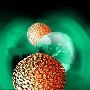
Murine norovirus with antibody fragments

![]()
Wall Art and Photo Gifts from Science Photo Library
Murine norovirus with antibody fragments
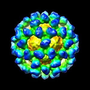
Murine norovirus (MNV) with antibody fragments, computer model. This image was created using molecular modelling software and data from cryo- electron microscopy. The image shows the outer protein shell of the virus (yellow, green), known as the capsid. It is covered with antibody fragments (light to dark blue) known as Fabs (antigen binding fragments). MNV belongs to the norovirus group of viruses, which are a major cause of non-bacterial gastroenteritis in humans. MNV infects only mice. Cryo-electron microscopy images specimens at a temperature of minus 150 degrees Celsius. It fires beams of electrons at multiple angles creating slices of data that are reconstructed into 3-D models on computer
Science Photo Library features Science and Medical images including photos and illustrations
Media ID 6412788
© THOMAS SMITH/DANFORTH CENTER/SCIENCE PHOTO LIBRARY
3 D Visualisation 3 D Visualization Antibody Capsid Computer Graphic Computer Rendering Cryo Electron Microscope Cryo Em Cryoelectron Microscopy Immune Response Immunology Macromolecular Macromolecule Modelling Molecular Imaging Noro Virus Particle Protein Shell Reconstruction Shape Structural Biology Surface Viral Shell Virion Virology Micro Biology Microbiological Molecular Model Molecular Structure Pathogen Protein Virus
FEATURES IN THESE COLLECTIONS
> Animals
> Fishes
> G
> Grouper
> Animals
> Insects
> Butterflies
> Smith Blue
EDITORS COMMENTS
This print showcases a computer-generated model of Murine norovirus (MNV) with antibody fragments, created using advanced molecular modelling software and data obtained from cryo-electron microscopy. The image highlights the outer protein shell of the virus, known as the capsid, depicted in vibrant shades of yellow and green. Fascinatingly, this capsid is adorned with antibody fragments, represented by varying tones of blue. MNV belongs to the notorious norovirus group responsible for causing non-bacterial gastroenteritis in humans. However, it exclusively infects mice rather than humans. Cryo-electron microscopy played a crucial role in capturing this stunning visual representation by imaging specimens at an incredibly frigid temperature of minus 150 degrees Celsius. By firing beams of electrons at multiple angles, slices of data were collected and subsequently reconstructed into intricate 3-D models on a computer. The significance of this image lies not only in its aesthetic appeal but also in its contribution to fields such as biology and medicine. It provides valuable insights into the structure and composition of MNV while shedding light on various aspects related to virology, immunology, and molecular imaging. Furthermore, it underscores the importance of structural biology research conducted through techniques like cryoelectron microscopy. This remarkable artwork serves as a testament to human ingenuity and our relentless pursuit to unravel nature's mysteries at a microscopic level.
MADE IN THE USA
Safe Shipping with 30 Day Money Back Guarantee
FREE PERSONALISATION*
We are proud to offer a range of customisation features including Personalised Captions, Color Filters and Picture Zoom Tools
SECURE PAYMENTS
We happily accept a wide range of payment options so you can pay for the things you need in the way that is most convenient for you
* Options may vary by product and licensing agreement. Zoomed Pictures can be adjusted in the Cart.