Home > Popular Themes > Red Arrows
Little and ring finger flexion, artwork C016 / 6793

![]()
Wall Art and Photo Gifts from Science Photo Library
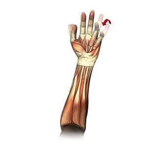
Little and ring finger flexion, artwork C016 / 6793
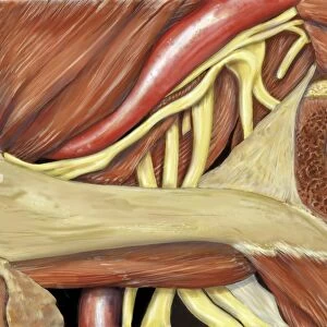
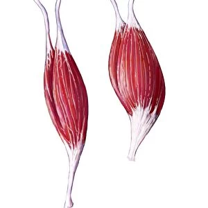
Little and ring finger flexion. Artwork of the muscles of the forearm and hand from the front, with red arrows showing the direction of movement of the little and ring (fourth and third) fingers during flexion. Finger flexion is controlled by the flexor digitorum superficialis, flexor digitorum profundus, and lumbrical muscles. Some muscles act on more than one finger. This is best demonstrated by flexing the little finger, causing involuntary flexion of the ring finger. This is because the ring and middle fingers, unlike the little and index fingers, lack independent extensor muscles to keep them extended as the other fingers flex. The nerve used is the ulnar nerve. This is the right hand. For the left hand, see C016/6794
Science Photo Library features Science and Medical images including photos and illustrations
Media ID 9243273
© D & L GRAPHICS / SCIENCE PHOTO LIBRARY
Anterior Arthrology Bending Biomechanics Coupled Diagram Finger Flexing Flexion Flexor Digitorum Profundus Flexor Digitorum Superficialis Forearm Frontal Hand Joint Joints Ligament Ligaments Limb Lumbrical Movement Moving Muscles Muscular Physiological Physiology Range Of Movements Tendon Tendons Ulnar Nerve Cutouts Little Finger Musculature Right Hand
FEATURES IN THESE COLLECTIONS
> Arts
> Art Movements
> Related Images
EDITORS COMMENTS
This print showcases the intricate movements of the little and ring fingers in the human hand. The artwork, titled "Little and ring finger flexion" delves into the muscles of the forearm and hand, highlighting their role in finger flexion. With red arrows indicating the direction of movement, this illustration sheds light on how these two fingers bend during flexion. The flexor digitorum superficialis, flexor digitorum profundus, and lumbrical muscles are responsible for controlling finger flexion. Interestingly, some muscles have an impact on multiple fingers. This is exemplified by the involuntary flexion of the ring finger when one attempts to solely move their little finger. Unlike the index and little fingers that possess independent extensor muscles to keep them extended during flexion, both middle and ring fingers lack such autonomy. In this image depicting a right hand against a white background, we gain insight into various aspects of anatomy including tendons, ligaments, musculature, joints, and nerves like the ulnar nerve involved in this process. It serves as a reminder that our bodies are marvelously complex systems with interdependent parts working together seamlessly. This detailed artwork not only provides valuable information about normal physiology but also highlights shared muscle functions between adjacent digits. A companion piece illustrating similar movements in a left hand is available as well (C016/6794). Overall, this print from D & L GRAPHICS / SCIENCE PHOTO LIBRARY offers an engaging exploration into biomechanics within our hands' intricate structures.
MADE IN THE USA
Safe Shipping with 30 Day Money Back Guarantee
FREE PERSONALISATION*
We are proud to offer a range of customisation features including Personalised Captions, Color Filters and Picture Zoom Tools
SECURE PAYMENTS
We happily accept a wide range of payment options so you can pay for the things you need in the way that is most convenient for you
* Options may vary by product and licensing agreement. Zoomed Pictures can be adjusted in the Cart.