Home > Animals > Mammals > Muridae > Water Mouse
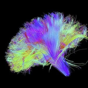
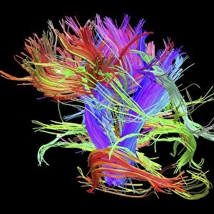
White matter fibres and brain, artwork C015 / 1930

![]()
Wall Art and Photo Gifts from Science Photo Library
White matter fibres and brain, artwork C015 / 1930
White matter fibres overlaid a 3d model of the human brain in top view. Coloured 3D diffusion spectral imaging (DSI) scan of the bundles of white matter nerve fibres in the brain. The fibres transmit nerve signals between brain regions and between the brain and the spinal cord. Diffusion spectrum imaging (DSI) is a variant of magnetic resonance imaging (MRI) in which a magnetic field maps the water contained in neuron fibers, thus mapping their criss-crossing patterns. A similar technique called diffusion tensor imaging (DTI) is also used to explore neural data of white matter fibres in the brain. Both methods allow mapping of their orientations and the connections between brain regions. Data/software: NIH Human Connectome Project /humanconnectomeproject.org)
Science Photo Library features Science and Medical images including photos and illustrations
Media ID 9241829
© PASIEKA/SCIENCE PHOTO LIBRARY
Connections Diffusion Spectral Imaging Diffusion Tensor Imaging Dsi Scan Dti Scan Fibers Fibre Tracking Fibres Human Brain Mapping Nerve Bundles Nerve Fibre Pathway Pathways White Matter Brain Connexions Nervous System
FEATURES IN THESE COLLECTIONS
> Animals
> Mammals
> Muridae
> Water Mouse
> Maps and Charts
> Related Images
EDITORS COMMENTS
This print showcases the intricate network of white matter fibres in the human brain, overlaid on a 3D model. The vibrant colors represent a 3D diffusion spectral imaging (DSI) scan, revealing the bundles of nerve fibres that transmit crucial signals between different regions of the brain and even to the spinal cord. By utilizing magnetic resonance imaging (MRI), DSI maps the water content within these neuron fibers, allowing for an accurate visualization of their criss-crossing patterns. The technique employed here is similar to diffusion tensor imaging (DTI), which also explores neural data related to white matter fibres. Both methods provide valuable insights into mapping their orientations and connections across various brain regions. This invaluable information aids researchers in understanding how different pathways are formed within our nervous system. The data used for this artwork originates from the NIH Human Connectome Project, an ambitious endeavor aimed at comprehensively mapping and understanding human brain connectivity. Through fiber tracking techniques like DTI and DSI scans, scientists can delve deeper into unraveling complex connexions within our brains. This mesmerizing image serves as a visual testament to the incredible complexity and beauty inherent in our most vital organ – the human brain. It offers us a glimpse into its inner workings while reminding us of how much there still is left to explore and understand about this remarkable masterpiece of nature's design.
MADE IN THE USA
Safe Shipping with 30 Day Money Back Guarantee
FREE PERSONALISATION*
We are proud to offer a range of customisation features including Personalised Captions, Color Filters and Picture Zoom Tools
SECURE PAYMENTS
We happily accept a wide range of payment options so you can pay for the things you need in the way that is most convenient for you
* Options may vary by product and licensing agreement. Zoomed Pictures can be adjusted in the Cart.