Home > Animals > Mammals > Muridae > Water Mouse
White matter fibres and brain, artwork C015 / 1939

![]()
Wall Art and Photo Gifts from Science Photo Library
White matter fibres and brain, artwork C015 / 1939
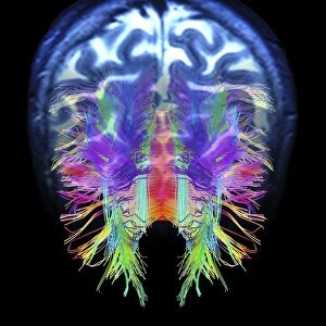
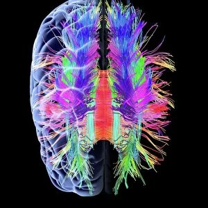
White matter fibres overlaid a 3d model of the human brain in top view. Coloured 3D diffusion spectral imaging (DSI) scan of the bundles of white matter nerve fibres in the brain. The fibres transmit nerve signals between brain regions and between the brain and the spinal cord. Diffusion spectrum imaging (DSI) is a variant of magnetic resonance imaging (MRI) in which a magnetic field maps the water contained in neuron fibers, thus mapping their criss-crossing patterns. A similar technique called diffusion tensor imaging (DTI) is also used to explore neural data of white matter fibres in the brain. Both methods allow mapping of their orientations and the connections between brain regions. Data/software: NIH Human Connectome Project /humanconnectomeproject.org)
Science Photo Library features Science and Medical images including photos and illustrations
Media ID 9212559
© PASIEKA/SCIENCE PHOTO LIBRARY
Connections Diffusion Spectral Imaging Diffusion Tensor Imaging Dsi Scan Dti Scan Fibers Fibre Tracking Fibres Human Brain Mapping Nerve Bundles Nerve Fibre Pathway Pathways White Matter Brain Connexions Nervous System
FEATURES IN THESE COLLECTIONS
> Animals
> Mammals
> Muridae
> Water Mouse
> Maps and Charts
> Related Images
EDITORS COMMENTS
This print showcases the intricate network of white matter fibres in the human brain, overlaid on a 3D model. The vibrant colors represent a 3D diffusion spectral imaging (DSI) scan, revealing the bundles of nerve fibres that transmit crucial signals between different regions of the brain and even to the spinal cord. Utilizing magnetic resonance imaging (MRI), this variant technique called diffusion spectrum imaging (DSI) employs a magnetic field to map water molecules within neuron fibers. This mapping allows for an exploration of their criss-crossing patterns, providing valuable insights into neural connectivity. Similar to DSI, diffusion tensor imaging (DTI) is another method utilized to study white matter fibres in the brain. Both techniques enable researchers to map orientations and connections between various brain regions with precision. The data used for this artwork originates from the NIH Human Connectome Project, which aims to comprehensively understand how different areas of our brains are interconnected. By visualizing these pathways through cut-outs and fibre tracking techniques, scientists can gain deeper knowledge about connexions within our nervous system. This mesmerizing image not only highlights the complexity and beauty of our neural architecture but also serves as a reminder of how far we have come in unraveling one of nature's greatest mysteries -the human brain.
MADE IN THE USA
Safe Shipping with 30 Day Money Back Guarantee
FREE PERSONALISATION*
We are proud to offer a range of customisation features including Personalised Captions, Color Filters and Picture Zoom Tools
SECURE PAYMENTS
We happily accept a wide range of payment options so you can pay for the things you need in the way that is most convenient for you
* Options may vary by product and licensing agreement. Zoomed Pictures can be adjusted in the Cart.