Home > Popular Themes > Human Body
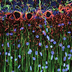
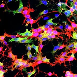
Cerebellum tissue, light micrograph

![]()
Wall Art and Photo Gifts from Science Photo Library
Cerebellum tissue, light micrograph
Cerebellum tissue. Confocal light micrograph of a section through the cerebellum of the brain. Purkinje cells, a type of neuron (nerve cell), are red. Radial glial cells, a type of support cell, are yellow, and cell nuclei are purple. Purkinje cells consist of a flask-shaped cell body with many branching processes (dendrites) that receive impulses from other cells. Purkinje cells form the junction between the granular and molecular layers of the grey matter of the cerebellum. The radial glial cells provide structural support, and nutrients and oxygen for the Purkinje cells. The cerebellum controls balance, posture and muscle coordination
Science Photo Library features Science and Medical images including photos and illustrations
Media ID 1698351
© C.J.GUERIN, PhD, MRC TOXICOLOGY UNIT/SCIENCE PHOTO LIBRARY
Central Nervous System Cerebellar Cerebellum Confocal Light Micrograph Dendrite Dendrites Fluorescence Fluorescent Glia Grey Matter Histological Histology Immunofluorescence Immunofluorescent Magnified Image Microscopic Subjects Nerve Cell Nervous Neuron Nuclei Nucleus Purkinje Cell Stain System Brain Cells Light Micrograph Light Microscope Neurological Neurology Section Sectioned
FEATURES IN THESE COLLECTIONS
> Science Photo Library
> Graphics and Patterns
EDITORS COMMENTS
This print showcases the intricate beauty of cerebellum tissue, as captured through a confocal light micrograph. The cerebellum, a vital part of the brain responsible for balance, posture, and muscle coordination, is depicted in stunning detail. Purkinje cells steal the spotlight with their vibrant red hue. These specialized neurons possess a unique flask-shaped cell body adorned with numerous branching processes called dendrites that receive signals from other cells. The granular and molecular layers of the cerebellar grey matter find their junction within these remarkable Purkinje cells. Supporting this delicate network are radial glial cells, portrayed in striking yellow tones. These support cells play an essential role by providing structural support to the Purkinje cells while also supplying them with nutrients and oxygen. Immersed in this microscopic world are countless purple-hued cell nuclei representing the core of cellular activity within this complex system. This magnified image offers insight into the intricate architecture and interconnectedness found within our central nervous system. With its blend of biology, anatomy, and neurology expertise on display, this histological masterpiece serves as a testament to both scientific discovery and artistic appreciation for nature's wonders.
MADE IN THE USA
Safe Shipping with 30 Day Money Back Guarantee
FREE PERSONALISATION*
We are proud to offer a range of customisation features including Personalised Captions, Color Filters and Picture Zoom Tools
SECURE PAYMENTS
We happily accept a wide range of payment options so you can pay for the things you need in the way that is most convenient for you
* Options may vary by product and licensing agreement. Zoomed Pictures can be adjusted in the Cart.