Home > Sport > Cycling
Cell division, fluorescent micrograph

![]()
Wall Art and Photo Gifts from Science Photo Library
Cell division, fluorescent micrograph
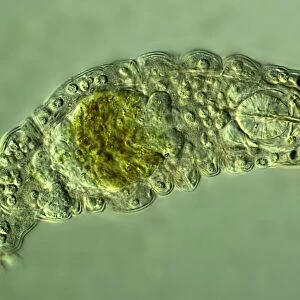
Cell division. Immunofluorescent light micrograph of a human epithelial cell (centre) during the late anaphase stage of mitosis. Mitosis is the cycle of replication and division by which new body cells are formed. Here the chromosomes (blue) of the parent cell have duplicated and the pairs are being pulled apart by microtubules (spindles, green) to opposite poles of the cell, in a process called cytokinesis. When the cell has split, two identical daughter cells are formed
Science Photo Library features Science and Medical images including photos and illustrations
Media ID 6324461
© DR TORSTEN WITTMANN/SCIENCE PHOTO LIBRARY
Cell Biology Cell Division Cellular Chromosome Chromosomes Cycle Cytokinesis Cytological Cytology Cytoplasm Daughter Cells Dividing Early Fiber Fibers Fibre Fibres Fluorescence Fluorescent Genetic Immunofluorescence Immunofluorescent Microtubule Microtubules Mitosis Mitotic Physiology Separating Separation Spindle Spindles Stage Stretching Genetics Light Micrograph Light Microscope Micro Biology
FEATURES IN THESE COLLECTIONS
EDITORS COMMENTS
This immunofluorescent light micrograph captures the late anaphase stage of mitosis in a human epithelial cell. Mitosis is a fundamental process in the life cycle of cells, enabling the formation of new body cells through replication and division. During mitosis, the chromosomes (blue) of the parent cell duplicate, and the pairs are pulled apart by microtubules (spindles, green) to opposite poles of the cell in a process called cytokinesis. In this image, the chromosomes have reached the maximum separation, and the cytoplasm (not visible) begins to pinch in between the separating chromosomes, eventually leading to the formation of two identical daughter cells. This physiological process is crucial for maintaining the health and integrity of tissues and organs in the human body. The microtubules, essential fibers for cell division, are highlighted in green, while the chromosomes are stained blue. This image showcases the intricacy and beauty of cellular processes, providing valuable insights into the field of cell biology, cytology, and genetics.
MADE IN THE USA
Safe Shipping with 30 Day Money Back Guarantee
FREE PERSONALISATION*
We are proud to offer a range of customisation features including Personalised Captions, Color Filters and Picture Zoom Tools
SECURE PAYMENTS
We happily accept a wide range of payment options so you can pay for the things you need in the way that is most convenient for you
* Options may vary by product and licensing agreement. Zoomed Pictures can be adjusted in the Cart.